
This article will give you a clear, evidence-based look at breast thermography. We’ll explore the science behind how it works, compare it directly to mammography, clarify the FDA’s official stance, and explain its appropriate role in a holistic wellness strategy. Our goal is to empower you with the facts so you can make informed decisions about your health.
Key Takeaways
- Adjunctive Use Only: The FDA has cleared thermography only as an additional tool to be used alongside a primary screening test like a mammogram, not as a standalone replacement.
- Functional vs. Structural: Thermography measures heat and blood flow patterns (function), while mammograms detect physical masses and calcifications (structure).
- No Proven Mortality Reduction: Mammography is the only screening method proven to reduce deaths from breast cancer through early detection.
- Not a Diagnostic Tool: An abnormal thermogram cannot be biopsied; it requires follow-up with structural imaging like a mammogram or ultrasound to investigate further.
- Holistic Insight: Thermography can offer valuable insights into physiological activity like inflammation, which can be useful within a broader, whole-body wellness plan.
Understanding Thermography: How Infrared Thermal Imaging Works
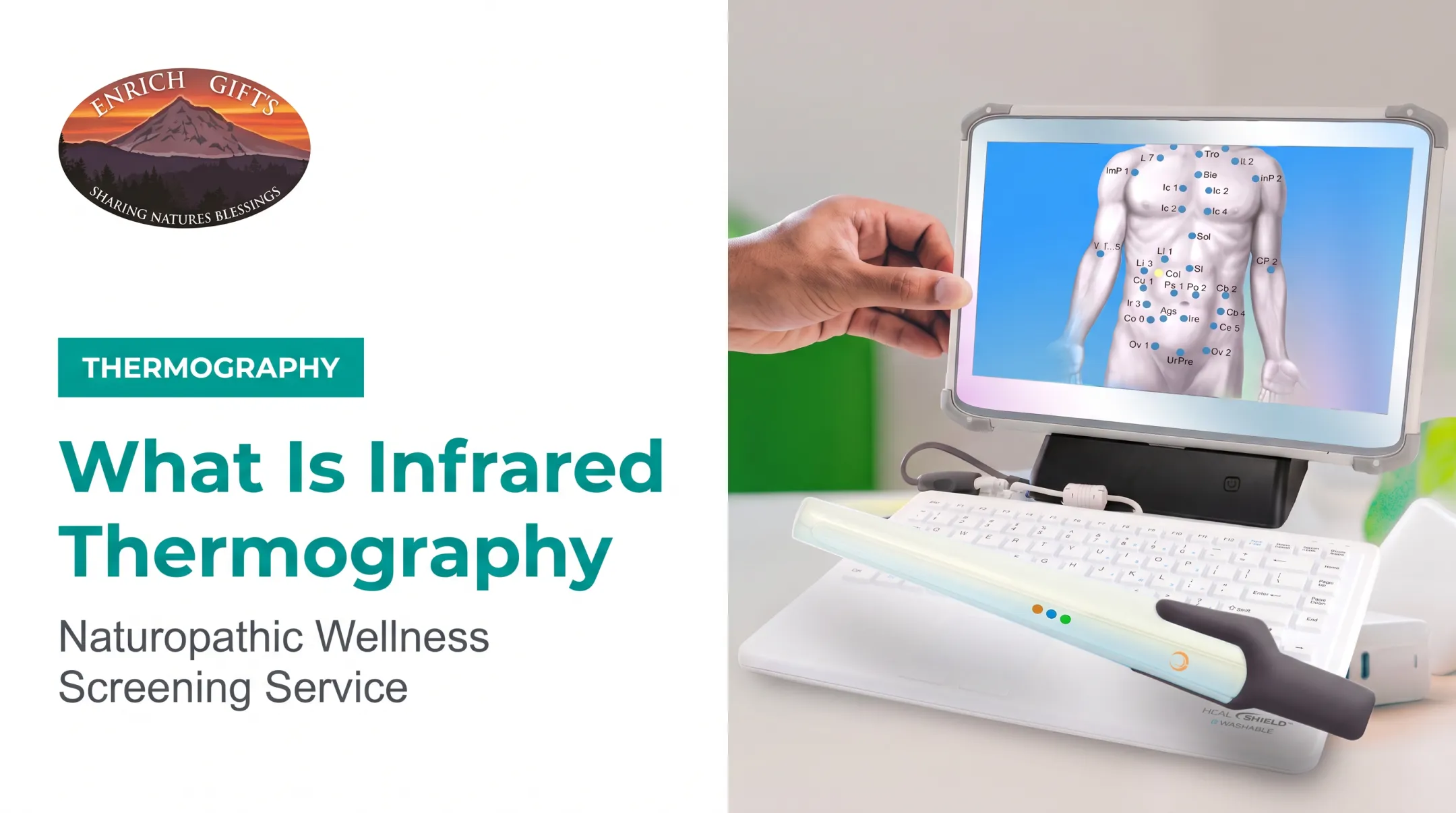
Digital Infrared Thermal Imaging (DITI), or thermography, is a non-invasive, no-touch assessment that uses a specialized infrared camera to map the temperature patterns on the surface of your skin. It doesn't emit radiation or compress tissue; it simply captures the heat your body naturally radiates.
Physiological processes generate heat. For breast health screening, thermography looks for thermal signals related to:
- Increased metabolic activity: Cancer cells can be more metabolically active than healthy cells, which may generate more heat.
- Angiogenesis (new blood vessel formation): Tumors often create their own blood supply to fuel their growth. This increased blood flow, or hypervascularity, can bring more heat to the skin’s surface.
- Inflammation: The body's inflammatory response also increases circulation and temperature.
Thermography measures physiological function. It maps heat patterns on the skin, but it does not image anatomical structures, tumors, or cellular changes inside the breast. Think of it as creating a "weather map" of your body's surface temperature.
Thermogram vs. Mammogram: Structural vs. Functional Imaging
Thermography and mammography are different technologies built for different jobs. Comparing them side by side clarifies what each test can and cannot do.
Key Differences in Technology and Purpose
A common point of confusion is thinking of these two tests as interchangeable. They are not.
- Mammogram: Uses low-dose X-rays to image internal breast tissue. Brief compression spreads the tissue so masses, lumps, and microcalcifications are easier to see.
- Thermogram: Uses an infrared sensor to map heat from the skin. It shows temperature differences and vascular patterns tied to physiological change.
One is looking at structure, the other at function. Compare it to inspecting a house. A mammogram is like reading the blueprints to find a misplaced wall. A thermogram is like using a thermal camera to find a hot spot that could signal faulty wiring.

Clinical Reliability and Evidence Base
When evaluating any screening test, the most important question is: does it save lives? This is where mammography has a long-established evidence base. The National Cancer Institute reports that mammography screening is associated with a relative reduction in breast cancer mortality of about 15-20% for women aged 50-69.
Thermography does not have this level of evidence. While it can detect heat, it hasn't been proven in large-scale clinical trials to reduce mortality from breast cancer when used as a primary screening tool.
Breast tissue density can also affect imaging. Dense tissue can hide abnormalities on a mammogram, so a doctor may add ultrasound or MRI.
The FDA states it knows of no scientific evidence supporting claims that thermography is a better cancer screening option for women with dense breasts.
The Diagnostic and Follow-up Pathway
The difference in follow-up procedures is also critical.
- Mammogram: A suspicious area can lead to more imaging and, if needed, a targeted needle biopsy guided to the exact structural finding.
- Thermogram: A hot spot or asymmetric pattern is a functional signal, not a mass, so there is nothing to biopsy. Next step is structural imaging, such as a mammogram or ultrasound, to look for a physical cause.
Clinical Reality: FDA Clearance, Detection Capabilities, and Limitations
Misleading marketing has fueled widespread confusion about what thermography can and cannot do. Knowing its actual regulatory status and limitations matters before relying on it for breast health screening.
The FDA's Regulatory Stance on Thermography
The U.S. Food and Drug Administration (FDA) maintains an unambiguous position on thermography's role. Thermography is cleared only as an "adjunctive" screening tool. This means it is cleared for use alongside a primary screening method like mammography, but never as a standalone test to screen for or diagnose breast cancer.
The FDA has issued warnings against clinics and websites that promote thermography as a substitute for mammography. In a 2019 safety communication, the agency specifically cautioned that relying solely on thermography could delay a cancer diagnosis and worsen health outcomes.
Understanding False Positives and False Negatives
Like any test, thermography carries risks of inaccurate results in both directions.
- False Negatives: A slow-growing or deep-seated tumor may not generate enough surface heat for the camera to detect, so the test misses a cancer that is actually present.
- False Positives: Benign conditions such as cysts, infections, or even recent vigorous exercise can raise skin temperature and trigger a false alarm.
A study of symptomatic patients published in Clinical Radiology found that DITI produced significant rates of both errors, a pattern that can cause unnecessary anxiety and lead to additional testing.

Quality Control and Regulatory Standards
Mammography is a highly regulated field. The Mammography Quality Standards Act (MQSA) requires facilities to be certified, technicians to be trained, and radiologists to be qualified. Equipment and radiation doses are strictly monitored.
The world of thermography has far more variable standards. While certifying bodies like the American College of Clinical Thermology exist, they are not part of a mandatory federal program like MQSA. The quality of the equipment, the training of the technician, and the expertise of the person interpreting the images can vary significantly from one provider to another.
What to Expect During a Breast Thermography Procedure
If you choose to have a breast thermogram as an adjunctive assessment, knowing what to expect can help ensure the results are as accurate as possible. The goal is to get a true baseline reading of your body's thermal patterns without interference.
Preparation and Protocol Before the Scan
To get an accurate thermal reading, you'll need to follow specific instructions. According to guidelines from the American Academy of Thermology, preparation may include:
- Skipping heavy exercise, yoga, or massage for at least 3 hours before the scan.
- Avoiding lotions, creams, powders, or deodorants on the chest or underarms on the day of the exam.
- Protecting skin from sun exposure or sunburn on the day of or day before the scan.
- Holding off on baths or hairdryer use within 1 hour of the test.
- Steering clear of smoking and alcohol for 4 hours prior.
The Step-by-Step Scanning Process
The procedure itself is simple and completely touchless.
- Acclimate: You'll enter a private, temperature-controlled room and disrobe from the waist up. You'll then sit or stand for about 15 minutes, allowing your body to adjust to the room's temperature.
- Capture Images: A trained technician will use a medical-grade infrared camera, positioned several feet away, to capture a series of images.
- Capture Multiple Views: The technician typically captures images from the front and both sides (oblique angles) for a complete picture of your thermal patterns. The entire process is painless and takes only a few minutes.
- Map the Data: The system sends captured data to a computer, which generates colorful images where different colors represent different temperatures.
Interpreting Thermal Symmetry and Baseline Tracking
Unlike a mammogram that looks for a specific mass, thermogram interpretation is often about symmetry and change over time. Your body's thermal pattern is unique to you. The primary goal of a first scan is to establish your personal "thermal fingerprint."
A follow-up scan, typically 3-6 months later, confirms that your baseline is stable. Once you establish a stable baseline, annual scans can help monitor for any significant asymmetric changes in temperature or vascular patterns that might warrant further investigation with your medical doctor.

Integrating Thermography into a Proactive Whole-Body Wellness Strategy
While thermography is not a substitute for mammography, it can be a valuable tool when placed in the right context: as part of a comprehensive, proactive approach to whole-body wellness.
Using Thermal Imaging for Functional Wellness Insights
Thermography can visualize inflammation and circulatory patterns beyond breast health alone. Practitioners use it to spot physiological stress and dysfunction throughout the body before symptoms or structural disease appear.
That insight supports targeted lifestyle and nutritional interventions—and helps answer, "Where is my body under stress?"
The Holistic Perspective with Enrich Gifts
At Enrich Gifts, our naturopath, Dr. Carolyn Johansen, uses functional assessments to help clients understand the root causes of their health concerns. We see the body as an interconnected system, and thermography is one of several tools we use to get a clearer picture of what's happening beneath the surface.
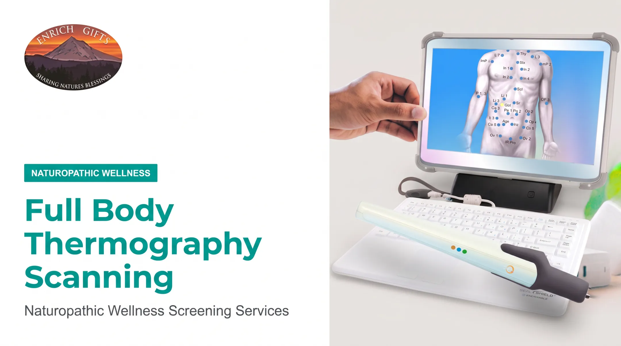
Dr. Johansen uses the AlfaSight 9000 thermography system to assess how major body systems are functioning. The focus is identifying patterns of inflammation, compensation, and dysfunction—not diagnosing disease. She then combines those findings with other assessments, like OligoScan (mineral imbalances and heavy metals) and metabolic evaluations, to build a personalized wellness plan.
Building a Comprehensive, Collaborative Health Plan
A proactive health strategy builds a collaborative team and draws on the best of both conventional and functional approaches.
This means:
- Continuing with proven screenings: Follow your doctor's recommendations for routine mammograms as the cornerstone of early breast cancer detection.
- Gaining functional insights: Use tools like thermography to monitor physiological function and flag areas for preventive support.
- Focusing on root causes: Work with a practitioner like Dr. Johansen on lifestyle, nutrition, and systemic inflammation for long-term health.
By combining these approaches, you can move beyond a reactive model of healthcare and take an empowered, informed role in your own well-being.
Frequently Asked Questions
How accurate is thermography in detecting breast cancer?
Thermography cannot reliably diagnose or rule out breast cancer on its own. It is not a validated standalone screening tool and can produce both false positives and false negatives, which is why the FDA has not cleared it as a substitute for mammography.
Is a thermogram better than a mammogram?
No, a thermogram is not better than a mammogram for breast cancer screening. A mammogram is the only imaging method that has been proven through extensive clinical trials to reduce mortality from breast cancer through early detection.
Does insurance cover thermography instead of a mammogram?
Most private health insurance plans and Medicare do not cover thermography for breast cancer screening because it is considered an adjunctive or investigational tool, not a primary screening method.
How much does a thermography scan cost?
Thermography is usually an out-of-pocket expense because most plans do not cover it. Price varies by provider and by whether you book a breast-only scan or a full-body assessment.
What should I avoid before a thermography scan?
Avoid anything that changes surface temperature before your scan: sun exposure, exercise, lotions or deodorants on the chest, smoking, and hot showers on the day of the appointment.
Which is better, thermography or ultrasound?
They serve different purposes. Ultrasound is a validated diagnostic tool for lumps or mammogram findings, especially in dense breast tissue; thermography is a non-diagnostic functional test that maps heat patterns.